Katalog
-
Katalog
- Auto & Motorrad
- Bauwesen und Immobilien
- Bekleidung
- Büro- und Schulartikel
- Chemikalien
- Dienstleistungen für Unternehmen
- Eisenwaren
- Elektrische Geräte & Zubehöre
- Elektronische Bauteile
- Energie
- Galanteriewaren
- Geschenke und Kunsthandwerke
- Gesundheit und Medizin
- Gummi und Kunststoffe
- Haus und Garten
- Haushaltsgeräte
- Koffer, Taschen & Hüllen
- Landwirtschaft
- Lebensmittel und Getränke
- Licht und Beleuchtung
- Maschinen, Geräte und Werkzeuge
- Maschinenteile und Herstellung Dienstleistungen
- Messapparat und Analysegerät
- Mineralien und Metallurgie
- Möbel
- Schuhe und Accessoires
- Schönheit und Körperpflege
- Service Geräte und -Ausstattung
- Sicherheit und Schutz
- Spielzeuge und Hobbys
- Sport und Unterhaltung
- Telekommunikations
- Textil und Lederware
- Transport
- Uhren, Schmuck, Brillen
- Umweltschutz
- Unterhaltungselektronik
- Verpacken und Drucken
- Werkzeuge
- Überschüssiger Warenbestand, Lager
Filters
Search
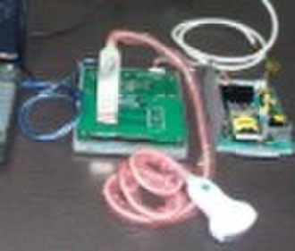
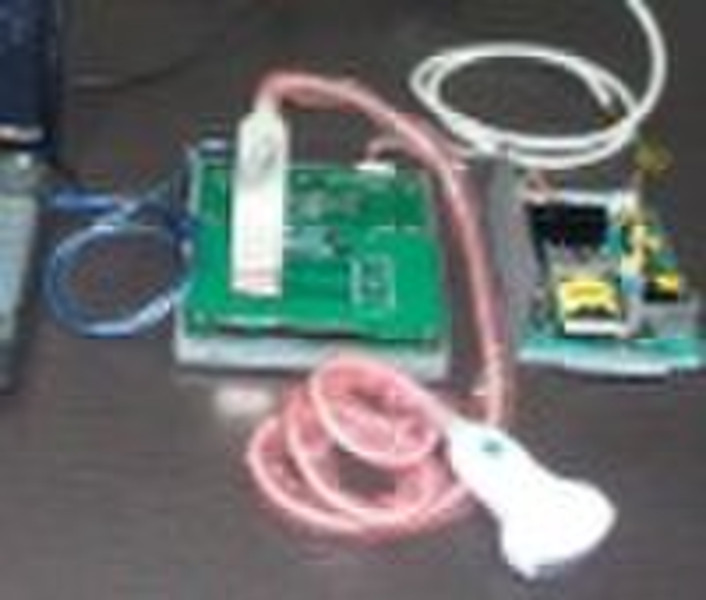
Ultraschall B Scanner Box
original-Preis: 1 000 USD
Shenzhen, China
Produktionskapazität:
500000 Einheit / Monat

Hill Lee
Kontaktperson
Basisdaten
| Ort der Herkunft | Guangdong China (Mainland) |
|---|---|
| Marke | Hurricane OEM |
| Modell-Nummer | Ultra-B |
When you use Ultrasound B Scanner Box, only need to connect to external 220V ac power, probe and any computer. Then install the software in the computer by the CD provide by us. Then can realizable the function of B Scanner, and at the same time use the other function of the computer, such as images manage and transport the images by internet.Parameters *Scanning depth: Maximum>=180mm. *Resolution: Lateral resolution: 4 Depth<=80mm, resolution<=2mm; 5 80mm<Depth<=130mm, resolution<=3mm; 6 130mm<Depth<=160mm, resolution<=4mm. Axial resolution: 2 Depth≤130mm, resolution≤1mm; 3 130mm<Depth≤170mm, resolution≤2mm. *Precision of geometry structure: Lateral: ≤10%; longitudinal: ≤5%. *Blind region: ≤3mm. *Transducer connector: ≥2; *Supported output apparatus: 2 USB devices; Digital printer, video printer, VCR *Monitor: 10 inch non-interlaced high-resolution monitor *Dimension: 25cm(height)*25cm (width)*45cm(length) *Weight: Around 16kg *Scanning mode: B, 2B, B/M, M, 4B, ZOOM; Real-time Zoom on B mode *Image gray scale: 256 level gray scale *Image processing: Pre-processing, after-processing, dynamic range, frame rate, line average, edge; enhancement, Black/White inversion; Gray scale adjustment, contrast, brightness, γ revision. *Gain: gain: adjustable between 0 100dB; Time gain control(TGC): 8 segment adjustment, B, M adjustment separately. *Cine loop: 64, 128, 256, 512frame, Auto/manual cine loop *Measurement and calculation: B mode: distance, circumstance, area, volume, angle, ratio, stenosis, profile, histogram; M mode: heart rate, time, distance, slope and stenosis; Gynecology measurement: Uterus, cervix, endometrium, L/R ovary; Obstetric: gestation age, fetal weight, AFI; Cardiology: LV, LV function, LVPW, RVAWT; Urology: transition zone volume, bladder volume, RUV, prostate, kidney; Small parts: optic, thyroid, jaw and face. *Storage: Image storage, cine loop, storage capacity≥80G. Measurement result and report can be stored. *Patient information management: Diagnosis case management, report print, comment library, image output (USB), import data. *Comments: comment, body mark(140 kinds for different parts), arrow. *Zoom: 10 ratio, 1.5, 2.0, 2.5, 3.0, 3.5, 4.0, 4.5, 5.0, 5.5, 6.0 *Multi-frequency: 5 segment frequencies *Scanning by angle: 12 angle scanning *Image direction: Up/down/left/right *Dynamic range(contrast): Continuous adjustable: 0 100dB. *Real-time Ethernet transmission(option): This option allows real-time transmission by Ethernet. Ethernet-capable allows local and internet ultrasound data transmission. *System preset: System preset includes parameter presets of OB, GYN, vascular, cardiac, urology and small parts, comments, manufacture default settings, system upgrade and maintenance settings. System configuration *Standard configuration: Ultrasound Box unit 1 Convex Probe 1 USB cable 1 Power cable 1 *Options: a) Transducers: Convex: R50, 2-5MHz, central frequency 3.5 MHz ; R60, 2-5MHz, central frequency 3.5MHz ; Micro-convex: R20, 2-5MHz, central frequency 3.0 MHz ; Linear : L40, 5-10MHz, central frequency 7.5 MHz ; Endocavity: R10, 4-9MHz, central frequency 6.5 MHz; Endocavity: R13, 4-9MHz, central frequency 6.5 MHz; Endocavity: R7, 4-9MHz, central frequency 6.5 MHz. b) Ultrasound report software: edit and print standard ultrasound report c) DICOM3.0, medical digital imaging and communication, is the industry standards (agreement) of image and data transmission among different kinds of medical devices. Ultrasound devices could receive images and data by DICOM after being connected to PACS. d) Cine loop(64, 128, 256, 512 frames) e) Real-time ethernet transmission f) Tissue harmonic imaging g) R/W CD room(This system is Not used) h) BW video printer i) Footswitch(freeze/ user-define) j) Maintenance tools k) Video apparatus
Lieferbedingungen und Verpackung
Packaging Detail: Standard Box Delivery Detail: 5-12days
Hafen: Shenzhen, HK
Zahlungsbedingungen
Letter of credit
Telegraphic transfer
Paypal
Western Union
-
Zahlungsarten
Wir akzeptieren:


