Katalog
-
Katalog
- Auto & Motorrad
- Bauwesen und Immobilien
- Bekleidung
- Büro- und Schulartikel
- Chemikalien
- Dienstleistungen für Unternehmen
- Eisenwaren
- Elektrische Geräte & Zubehöre
- Elektronische Bauteile
- Energie
- Galanteriewaren
- Geschenke und Kunsthandwerke
- Gesundheit und Medizin
- Gummi und Kunststoffe
- Haus und Garten
- Haushaltsgeräte
- Koffer, Taschen & Hüllen
- Landwirtschaft
- Lebensmittel und Getränke
- Licht und Beleuchtung
- Maschinen, Geräte und Werkzeuge
- Maschinenteile und Herstellung Dienstleistungen
- Messapparat und Analysegerät
- Mineralien und Metallurgie
- Möbel
- Schuhe und Accessoires
- Schönheit und Körperpflege
- Service Geräte und -Ausstattung
- Sicherheit und Schutz
- Spielzeuge und Hobbys
- Sport und Unterhaltung
- Telekommunikations
- Textil und Lederware
- Transport
- Uhren, Schmuck, Brillen
- Umweltschutz
- Unterhaltungselektronik
- Verpacken und Drucken
- Werkzeuge
- Überschüssiger Warenbestand, Lager
Filters
Search
BIR 500/225
original-Preis: 40,00 USD
Peking, China
Produktionskapazität:
20 Einstellen / Jahr
Peking, China
0086-10-64987563
64913897
64913897

ShengKam Yu
Kontaktperson
Basisdaten
| Ort der Herkunft | Illinois United States |
|---|---|
| Marke | ACTIS |
| Modell-Nummer | BIR 500/225 |
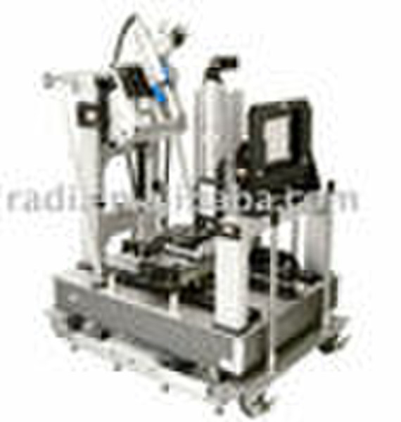
Introduction: BIR 500/225 FP is a combined Computed Tomography (CT) and Digital Radiography (DR)system designed to meet stringent requirements for spatial resolution, contrast discrimination, precision,repeatability, long term stability, and reliability. The system comprises the following components andsubsystems:• Comet 225 kV “twin-head” microfocus x-ray system with directional and transmission targets.Focus sizes of 6 μm directional and 2 μm transmission, with detection capability of 2 μm and 0.5 μmrespectively.• Varian PaxScan 4030E amorphous silicon flat panel detector with 3200 x 2304 pixels at 127 μm• Precision servo controlled multi-axis part manipulator with turntable, 50 cm vertical travel, 40 cmX axis translation, and 40 cm Y axis magnification.• 1.22 m x 1.83 m optical grade vibration damped mounting table.• Quad Core Intel®Xeon™ workstation, software, and flat panel color image displays;• Windows XP OS and ACTIS application software.• Integration, testing, installation, training, documentation, and one year warrantySystem Description BIR 500/225 FP is a Volume Computed Tomography (VCT) system with 2D digital radiography and “live” x-ray imaging capability. The system consists of a 225 kV Comet FXE-225.99 “twin-head” µfocus x-ray unit, Varian PaxScan 4030E amorphous silicon flat panel detector, quad core Xeon workstation, detector interface, four-axis part manipulator with linear servo drives, variable source-object distance, color image display, software, cabling, accessories, and documentation. The system is suitable for x-ray inspection of metallic and non-metallic items over a wide range of density and materials. The system produces cross sectional CT images, cone beam VCT images, in-motion “live” images and digital radiographs (DR). In addition to density mapping, CT provides complete 3-D morphology of parts with highly accurate dimensioning capability. CT also provides superior flaw detection capability of extremely small cracks, porosity and voids that are not visible with film radiography or RTR inspection.3. Object Size & Weight -500 mm diameter, 25 kg; PaxScan 4030E 40 to 225 kV Gigabit Ethernet; 3200 x 2304; 127 micron x 127 micron; 40.64 cm x 29.26 cm; 3 Hz at full resolution or 15 Hz with 2x2 binning; < 1% after gain and offset corrections (always applied); < 6% after first frame; 635 x 330 x 32 mm; 8000:1 (78 db). Object height limited only by cabinet interior dimensions;Maximum CT field of view is 450 mm diameter.4. CT Image Resolution -True Spatial Resolution (lines & spaces in gauge are resolved)2 µm for objects up to 5 mm diameter (2 µm focus, transmission target);5 µm for objects up to 10 mm diameter (2 µm focus, transmission target);10 µm for objects up to 20 mm diameter (6 µm focus, directional target);20 µm for objects up to 40 mm diameter (6 µm focus, directional target);At larger diameters, achievable spatial resolution will be ≤ (Field of View)/2500.Detail Detection (minimum detectable feature size)1 µm for objects up to 5 mm diameter (2 µm focus, transmission target);2.5 µm for objects up to 10 mm diameter (2 µm focus, transmission target);5 µm for objects up to 20 mm diameter (6 µm focus, directional target);10 µm for objects up to 40 mm diameter (6 µm focus, directional target);At larger diameters, achievable detail detection will be ≤ (Field of View)/5000.8. Pixel Size in Object - 1 µm to 1 mm depending on object diameter and matrix size. CT image field of view maximum sizeis 500 mm in ARO scanning mode.
Zahlungsbedingungen
Documents Against Acceptance
Documents Against Payment
Letter of credit
Telegraphic transfer
-
Zahlungsarten
Wir akzeptieren:


